
Pearson’s new 10th edition (1292026375, 9781292026374) and Marieb & Smith’s manuals offer extensive instructor support, aiding lab quizzing and exploration.
Importance of Hands-on Learning
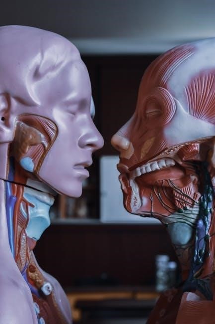
Hands-on laboratory experiences are crucial for solidifying theoretical knowledge in anatomy and physiology. Simply reading about complex biological systems isn’t enough; students truly grasp concepts through direct observation and manipulation. Laboratory manuals, like those from Pearson and Marieb, facilitate this by providing structured exercises – dissections, microscopy, and physiological measurements – that bring textbook material to life.
These practical sessions enhance critical thinking, problem-solving skills, and the ability to apply scientific methods. Furthermore, labs foster a deeper understanding of anatomical structures and physiological processes, moving beyond rote memorization. Accessing resources, even in PDF format, supports independent study and reinforces learning outside the scheduled lab time, ultimately preparing students for future healthcare professions.
Popular Manuals: Pearson & Marieb
Pearson and Marieb & Smith consistently rank among the most widely used anatomy & physiology laboratory manuals. Pearson’s 10th edition (ISBN: 1292026375, 9781292026374) is noted for its comprehensive coverage and updated exercises. Marieb’s manuals, often found as PDF versions through various online sources, are praised for their clear illustrations and step-by-step instructions.
Both publishers offer accompanying resources for instructors, including quizzes and assessment materials. These manuals typically cover a broad range of topics, from basic anatomical terminology and microscopy to detailed dissections of organ systems. Students benefit from the structured approach, enabling them to effectively translate theoretical knowledge into practical skills within the laboratory setting.
Accessing PDF Versions & Copyright Considerations
PDF versions of anatomy & physiology laboratory manuals, like those by Marieb & Smith, are frequently sought online. However, accessing these materials requires careful consideration of copyright laws. While some institutions provide legitimate access to students, downloading from unauthorized sources constitutes copyright infringement.
Websites offering free PDF downloads may contain outdated or inaccurate information, potentially hindering learning. Supporting publishers by purchasing authorized copies ensures continued development of high-quality educational resources. The Internet Archive actively archives web pages, but access to copyrighted materials remains restricted. Respecting intellectual property is crucial for maintaining academic integrity and supporting the educational community.
Core Concepts Covered in A&P Labs
A&P labs delve into anatomical terminology, microscopy, and histology, building a foundational understanding of the human body’s structure and function.
The Language of Anatomy: Anatomical Terminology
Understanding anatomical language is crucial for precise communication in A&P studies. Laboratory manuals, like those by Pearson and Marieb, dedicate sections to mastering directional terms – superior, inferior, anterior, posterior, medial, and lateral – establishing a framework for describing body locations.
Regional terms, denoting specific body areas (e;g., cephalic, thoracic, abdominal), are also emphasized. Students learn to apply these terms during dissections and when identifying structures under a microscope.
Furthermore, the manuals introduce planes of the body – sagittal, frontal (coronal), and transverse – essential for visualizing anatomical sections. Proficiency in this “language” is fundamental for interpreting lab results and advancing in anatomical studies.
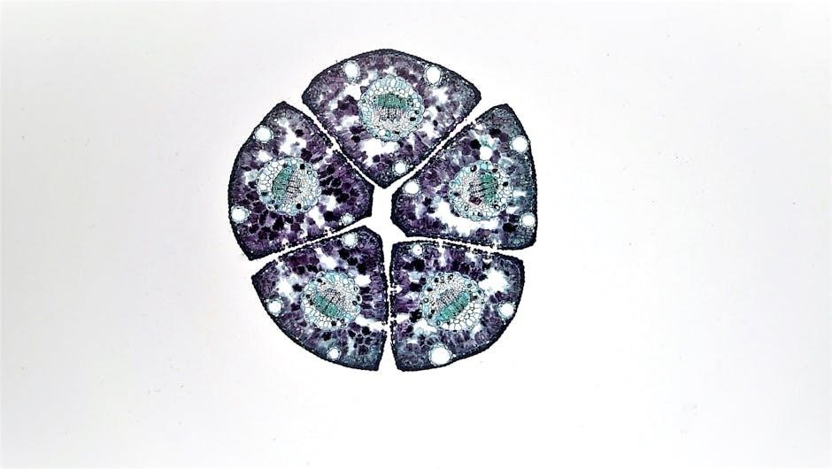
Microscopy Techniques & Histology
A&P laboratory manuals heavily emphasize microscopy, a cornerstone of histological study. Students learn proper microscope usage, including focusing techniques and objective lens selection, to observe tissues at a cellular level. Manuals detail slide preparation methods – staining, mounting – crucial for visualization.
Histology labs focus on identifying the four primary tissue types: epithelial, connective, muscle, and nervous.
Students practice differentiating these tissues based on their structure and function, often using prepared slides. Understanding staining techniques (like H&E) is vital for interpreting histological features. These skills are foundational for understanding organ structure and disease processes.
Essential Laboratory Equipment
A&P labs require microscopes for histological studies and dissection tools for anatomical exploration, alongside safety equipment ensuring a secure learning environment for students.
Microscope Usage & Maintenance
Microscopy is fundamental in A&P labs, enabling visualization of cells and tissues. Proper usage begins with correct slide preparation and focusing techniques, starting with low power objectives.
Understanding magnification and resolution is crucial for accurate observation. Regular maintenance, including cleaning lenses with appropriate solutions and proper storage, extends the microscope’s lifespan.
Students must learn to identify microscope parts – ocular lens, objective lenses, stage, and illumination controls – and troubleshoot common issues like blurry images. Careful handling prevents damage to this essential laboratory instrument, ensuring reliable histological analysis.
Dissection Tools & Safety Protocols
Successful dissections require appropriate tools – scalpels, forceps, scissors, and probes – used with precision and care. Prior to any dissection, a thorough understanding of safety protocols is paramount.
Students must wear gloves and eye protection to prevent exposure to specimens and potential hazards. Proper handling and disposal of dissection waste are essential for maintaining a safe laboratory environment.
Respectful treatment of specimens and adherence to instructor guidelines are non-negotiable. Familiarity with emergency procedures, including first aid for cuts, ensures a secure learning experience.
Human Tissue Examination
Laboratory manuals facilitate identifying epithelial and connective tissues, understanding their diverse types and crucial functions within the human body’s complex organization.
Epithelial Tissue Identification
Epithelial tissues, covering body surfaces and lining cavities, are categorized by cell shape (squamous, cuboidal, columnar) and layering (simple, stratified, pseudostratified).
Laboratory exercises focus on microscopic observation of prepared slides, enabling students to differentiate these types. Identifying key features like nuclei position, cell junctions, and presence of cilia is crucial.
Understanding the correlation between structure and function is paramount; for example, simple squamous epithelium facilitates diffusion in the lungs, while stratified squamous epithelium provides protection in the skin.
Manuals often include detailed diagrams and descriptions to aid in accurate identification, fostering a strong foundation for further histological studies.
Connective Tissue Types & Functions
Connective tissues – including connective tissue proper, cartilage, bone, and blood – provide support, protection, and connection throughout the body. Laboratory investigations emphasize recognizing diverse tissue types under the microscope.
Students learn to distinguish between dense and loose connective tissues, identifying components like collagen and elastin fibers. Cartilage types (hyaline, elastic, fibrocartilage) are differentiated based on matrix composition.
Bone tissue examination reveals osteons and canaliculi, while blood smears showcase erythrocytes, leukocytes, and platelets.
Understanding how structure dictates function – for instance, the cushioning role of cartilage or the rigidity of bone – is key to mastering connective tissue histology.

Skeletal System Laboratory Activities
Labs focus on bone identification, anatomical features, and joint classifications, exploring movement types and skeletal system functionality through practical dissections.
Bone Identification & Anatomy
Skeletal system labs heavily emphasize accurate bone identification, requiring students to distinguish various bones based on their unique morphological characteristics. This involves recognizing key anatomical landmarks – processes, foramina, tubercles, and fossae – that serve as attachment points for muscles, ligaments, and tendons.
Students will learn to classify bones by shape – long, short, flat, irregular, and sesamoid – and correlate bone structure with its specific function within the body. Practical exercises often include assembling skeletal models, disarticulating and articulating bones, and identifying features on skeletal preparations.
Understanding bone anatomy is crucial for comprehending biomechanics, movement, and the overall structural support provided by the skeletal system. Detailed observation and careful labeling are essential components of these laboratory activities.
Joint Classification & Movement
Laboratory activities focusing on joint classification involve categorizing joints based on their structural composition – fibrous, cartilaginous, and synovial – and their functional properties – synarthrosis, amphiarthrosis, and diarthrosis. Students will examine models and diagrams to identify the key features of each joint type.
A significant portion of these labs is dedicated to exploring the range of motion permitted by different synovial joints – hinge, pivot, ball-and-socket, condylar, saddle, and plane. Students will actively demonstrate and analyze movements like flexion, extension, abduction, adduction, rotation, and circumduction.
Understanding the relationship between joint structure and movement is vital for comprehending musculoskeletal function and potential pathologies.
Muscular System Dissections & Studies
Labs explore muscle tissue types – skeletal, smooth, and cardiac – focusing on their unique characteristics and functions through dissections and microscopic examination.
Muscle Tissue Types & Characteristics
Anatomy & Physiology laboratory studies delve into the three primary muscle tissue types: skeletal, smooth, and cardiac. Skeletal muscle, responsible for voluntary movements, exhibits striated appearance under microscopy and is attached to bones via tendons. Smooth muscle, found in the walls of internal organs, lacks striations and controls involuntary functions like digestion.
Cardiac muscle, exclusive to the heart, displays striations and involuntary control, ensuring rhythmic contractions. Lab exercises involve microscopic observation to differentiate these tissues based on cellular structure, fiber arrangement, and presence or absence of striations. Understanding these characteristics is crucial for comprehending muscle function and overall physiological processes.
Muscle Anatomy & Action
Laboratory explorations focus on identifying major skeletal muscles, their origins, insertions, and actions. Dissections and anatomical models reveal muscle fiber arrangements – parallel, pennate, and circular – influencing force production. Students learn to correlate muscle names with their functions, such as biceps brachii for elbow flexion and gastrocnemius for plantar flexion.
Physiological studies examine muscle contraction mechanisms, including the sliding filament theory. Analyzing muscle leverage and joint angles demonstrates how muscles generate movement. Understanding antagonistic muscle pairs – like flexors and extensors – is vital for comprehending coordinated body movements and overall musculoskeletal system functionality.
Nervous System Exploration
Labs involve brain dissections to identify structures and spinal cord anatomy studies, alongside reflex arc investigations, enhancing neurological pathway comprehension.
Brain Dissection & Structure
Brain dissection provides invaluable hands-on experience, allowing students to directly visualize the complex structures of the central nervous system. Laboratory manuals, like those by Pearson and Marieb, guide students through identifying key anatomical features, including the cerebrum, cerebellum, and brainstem.
Careful dissection reveals the gyri and sulci, responsible for increasing surface area, and allows for observation of the different lobes – frontal, parietal, temporal, and occipital – each associated with specific functions. Students learn to locate critical structures like the hypothalamus, thalamus, and medulla oblongata, understanding their roles in regulating vital processes.
This practical approach solidifies theoretical knowledge, fostering a deeper understanding of neurological pathways and the brain’s intricate organization.
Spinal Cord Anatomy & Reflexes
Laboratory exercises focusing on the spinal cord build upon brain studies, revealing the central nervous system’s continuous pathway. Manuals detail identifying the dorsal and ventral horns, the gray and white matter, and the protective meningeal layers through dissection and models.
Students investigate the organization of spinal nerves and their corresponding dermatomes, understanding how specific regions of the body are innervated. A crucial component involves exploring reflexes – specifically, testing and analyzing reflex arcs like the patellar reflex.
This hands-on approach demonstrates the speed and efficiency of neural pathways, solidifying comprehension of sensory and motor functions controlled by the spinal cord.
Cardiovascular System Investigations
Labs involve heart dissection to trace blood flow, alongside blood analysis examining components. Manuals aid understanding of circulatory pathways and physiological functions.
Heart Dissection & Blood Flow
Heart dissection provides a crucial hands-on experience, allowing students to visualize the chambers, valves, and major vessels responsible for circulating blood throughout the body. Laboratory manuals, like those by Pearson and Marieb, guide this process, detailing the correct techniques for safe and effective dissection.
Students trace the pathway of blood flow, starting with deoxygenated blood entering the right atrium and following it through the pulmonary circuit to the lungs and back to the left side of the heart. Understanding this flow is paramount.
Furthermore, investigations often include observing the heart’s structure in relation to its function, identifying key anatomical features, and correlating these with physiological principles. This reinforces comprehension of cardiovascular dynamics.
Blood Analysis & Components
Blood analysis is a cornerstone of A&P labs, enabling students to investigate the composition and function of this vital fluid. Manuals, such as those from Pearson and Marieb, detail procedures for hematocrit determination, blood typing, and differential white blood cell counts.
Labs commonly involve microscopic examination of blood smears to identify erythrocytes, leukocytes, and thrombocytes, understanding their respective roles in oxygen transport, immunity, and coagulation.
Students learn to interpret results, correlating them with physiological states and potential pathologies. This practical experience solidifies understanding of blood’s complex components and its critical role in maintaining homeostasis within the body.